185 West End Ave, Lincoln Towers, New York, NY 10023 | Telephone 212.877.3062
Diagnostic Ultrasound & Orthotic Scanning
Diagnostic Ultrasound
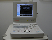
 At American Foot Care, Dr. Antonetz uses diagnostic ultrasound for high resolution imaging as an exam for injuries, soft tissue conditions of your foot and ankle. Ultrasound produces images of internal structures through the use of high-frequency sound waves, whose echoes are used to create moving and still images of the muscles, tendons, ligaments, joints and other soft tissue in the foot and ankle.
Dr. Antonetz may perform ultrasound to determine the cause of your pain. Many conditions can be visualized and diagnosed, such as: Achilles tendonitis, bursitis, plantar fasciitis, heel spurs, Morton's neuroma, stress fractures, joint and tendon injuries, and a wide range of other conditions. The images produced during this exam can be viewed in real time on a monitor during the procedure, and can produce 3-D images.
Benefits of Ultrasound
Diagnostic ultrasound is a safe, painless and can be performed in Dr. Antonetz's office. It can be used to effectively visualize many types of soft tissues and injuries that do not show up well during x-ray exams. The real-time images produced during an ultrasound exam also allow it to be used for guidance of surgical procedures if needed, which will ensure the most precise results.
There is no ionizing radiation used during the ultrasound procedure, making it safe for nearly all patients with no known risks or side effects.
Ultrasound Procedure
The ultrasound procedure begins with the patient lying down on the examination table as a water-based gel is applied to the area on their body that will be observed. This gel allows consistent contact between the body and the ultrasound transducer. The transducer is kept firmly against the skin and is moved over and across the area to allow for the most detailed visualization possible. The whole procedure usually takes 30 minutes.
There is no pain or discomfort associated with this procedure, however if the part of your foot being examined has already been injured there may be some slight pressure felt against it. If a Doppler type ultrasound is used, you may actually hear the pulses of your blood flow. There is no clinical risk inherent in ultrasonography as it uses no invasive methods, no ionizing radiation, and does not cause any health problems.
To learn more about our diagnostic ultrasound procedure, please call us today to schedule an appointment.
Digital Orthotic Scanning
 Foot-Orthotics are customized shoe inserts that correct abnormal foot movement and structural deformities during movement and rest. They provide comfort and functionality for people with various foot, ankle, knee, hip and back conditions. Historically, custom orthotics were typically made by creating a cast of the foot out of plaster. The orthotic was then designed using this plaster model of the foot.
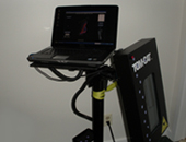
However, advances in technology now allow for an even more accurate and precise 3-D digital model of your foot. Digital orthotic scanning provides a more thorough assessment of your foot. To begin the digital orthotic scanning process, Dr. Antonetz places your foot held in the ideal corrected position against the Tom Cat Scanner™. The imaging creates from the scanner a 3-D map of the foot, which effectively images the biomechanical structure and any foot abnormalities. This information is digitally sent directly to the orthotic lab.
Compared with traditional casting methods used for creating orthotics, digital orthotic scanning offers a wide variety of benefits to both doctors and patients. Some of these benefits include:
- Speed - the results of digital orthotic scanning are processed immediately, unlike a plaster cast which usually has to dry first then be shipped to the orthotic lab
- Precision - digital orthotic scanning provides precise results, resulting in improved patient satisfaction and a reduced need for orthotic device adjustment
- Reporting - each digital orthotic scanning session creates a report with detailed results that cannot be matched by traditional methods
Digital orthotic scanning also provides faster analysis and production times than a plaster cast. Patients can receive their orthotics and start enjoying the comfort and stability much quicker. Dr. Antonetz is proud to offer this advanced technology to his patients.
Call our office today and schedule a consultation to see how you may benefit from digital orthotic scanning.


